| Center for Breast Health at Baptist for Women | |
|---|---|
| Phone: | 601-973-3180 |
| Fax: | 601-973-1601 |
Located off North State Street our entrance is between two large pink ribbons. | |

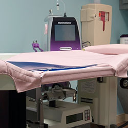
Mammography is a specific type of imaging that uses a low-dose x-ray system to examine breasts. A mammography exam, called a mammogram, is used to aid in the early detection and diagnosis of breast diseases in women.
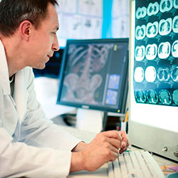
An x-ray (radiograph) is a noninvasive medical test that helps physicians diagnose and treat medical conditions. Imaging with x-rays involves exposing a part of the body to a small dose of ionizing radiation to produce pictures of the inside of the body. X-rays are the oldest and most frequently used form of medical imaging.
Three recent advances in mammography include digital mammography, computer-aided detection and breast tomosynthesis.
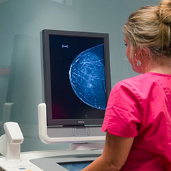
Digital mammography, also called full-field digital mammography (FFDM), is a mammography system in which the x-ray film is replaced by solid-state detectors that convert x-rays into electrical signals. These detectors are similar to those found in digital cameras. The electrical signals are used to produce images of the breast that can be seen on a computer screen or printed on special film similar to conventional mammograms. From the patient’s point of view, having a digital mammogram is essentially the same as having a conventional film mammogram.
Computer-aided detection (CAD) systems use a digitized mammographic image that can be obtained from either a conventional film mammogram or a digitally acquired mammogram. The computer software then searches for abnormal areas of density, mass, or calcification that may indicate the presence of cancer. The CAD system highlights these areas on the images, alerting the radiologist to the need for further analysis.
Women should have a mammogram once a year starting at the age of 40. The risk of breast cancer does go up with age – in fact the two main risk factors are gender and getting older. Mammograms should not be the only test for examining your breast. Self exams are also extremely important.
If you have a personal physician, she or he can provide you with a referral to the Center for Breast Health. You can also self-refer IF you have seen your referring physician in the past two years. We will ensure that your physician receives your results and any information for an appropriate follow-up.
Studies have shown digital mammography improves detection in women with dense breast tissue, those under the age of 50 and those in pre- or post-menopause. Digital mammography offers higher-contrast images that improbe detection. Instead of film, images are displayed on a computer monitor where they can be enhanced or magnified for a closer look.
The procedure is the same as for conventional mammography, but the new technology allows a quicker exam with a lower dose of radiation. Because the images are immediately available, there is less need to call women back for additional views, and the images can be sent instantly to the referring physician.

These procedures may be necessary if a mammogram or ultrasound detects something that needs further evaluation to provide an accurate diagnosis. In these procedures, no general anesthesia is necessary, only local anesthesia. A radiologist and a technologist are involved with you during this procedure. Several breast tissue samples are extracted. The procedure is usually less than 2 hours and the patients is able to go home.

Bone density scanning, also called dual-energy x-ray absorptiometry (DXA) or bone densitometry, is an enhanced form of x-ray technology that is used to measure bone loss. DXA is today’s established standard for measuring bone mineral density (BMD).
An x-ray (radiograph) is a noninvasive medical test that helps physicians diagnose and treat medical conditions. Imaging with x-rays involves exposing a part of the body to a small dose of ionizing radiation to produce pictures of the inside of the body. X-rays are the oldest and most frequently used form of medical imaging.
DXA is most often performed on the lower spine and hips. In children and some adults, the whole body is sometimes scanned. Peripheral devices that use x-ray or ultrasound are sometimes used to screen for low bone mass. In some communities, a CT scan with special software can also be used to diagnose or monitor low bone mass (QCT). This is accurate but less commonly used than DXA scanning.

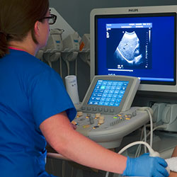
Ultrasound imaging, also called ultrasound scanning or sonography, involves exposing part of the body to high-frequency sound waves to produce pictures of the inside of the body. Ultrasound exams do not use ionizing radiation (x-ray). Because ultrasound images are captured in real-time, they can show the structure and movement of the body’s internal organs, as well as blood flowing through blood vessels.
Ultrasound imaging is usually a painless medical test that helps physicians diagnose and treat medical conditions.
Conventional ultrasound displays the images in thin, flat sections of the body. Advancements in ultrasound technology include three-dimensional (3-D) ultrasound that formats the sound wave data into 3-D images. Four-dimensional (4-D) ultrasound is 3-D ultrasound in motion.
A Doppler ultrasound study may be part of an ultrasound examination.
Doppler ultrasound is a special ultrasound technique that evaluates blood as it flows through a blood vessel, including the body’s major arteries and veins in the abdomen, arms, legs and neck.
For more information on ultrasound please visit radiologyinfo.org.
You need to bring your insurance card and driver’s license. You will not need a driver. You will not receive any medication.
Abdomen or gallbladder ultrasound: Do not eat or drink after midnight (NPO), including no smoking or gum chewing. You may brush your teeth.
Pelvic Ultrasound: Do not go to the bathroom 2 hours before the exam. During the first hour before the exam, you need to drink one 8 ounce glass of clear liquid every 15 minutes. It can be any clear liquid (Kool-aid, coffee, coke, etc. Examples of nonclear liquids are milk or juice.) Stop drinking 1 hour before exam.
For example: If appointment time is 2:00 PM, do not go to the bathroom after 12:00 noon. Starting at 12 noon until 1:00 PM, drink one glass of water every 15 minutes. Stop drinking the water at 1:00 PM. Patient may come in early to complete paperwork, 15 minutes is usually enough time to complete the paperwork.
Abdomen and pelvic ultrasound: Do not eat after midnight. NO FOOD. Drink one 8 ounce glass of water every 15 minutes beginning 2 hours before appointment time. This will be a total of four 8 ounce glasses (32 ounces). Stop drinking one hour before your appointment time.
Renal ultrasound: You may eat or drink with no restrictions. Do not go to the bathroom one hour before the examination. You do not need to force fluids, just have a normally full bladder until the examination is completed.
All other types of ultrasounds: There are no preparations for other ultrasound examinations except for biopsies.
You will be asked to remove most of your clothing and instructed to put on a gown. Sonograms require a gel be applied to your skin. This is so our transducer will glide across your skin and to insure there is contact between your skin and the transducer.
Follow the directions given for the location you are visiting found on our location pages. Upon arriving, check in with the receptionist who may have information for you to fill out. These forms are also available online. If you wish to fill them out ahead of time click here.
Once all your information has been obtained and processed, your technologist is notified of your arrival.
You will lie on an examination table. A warm gel will be applied to your skin, then a small instrument called a transducer will be moved across your skin. The transducer sends sound waves into your body. These high frequency sound waves are reflected back to the transducer. This information is processed by the ultrasound machine in real time producing sonographic images of the inside of your body. You cannot hear the sounds and the study is generally painless.
If you are female and are having a pelvic ultrasound, an endovaginal scan may be performed. This may be a little uncomfortable but should not be painful.
The most important thing is for you to follow the instructions for the preparation before the sonogram. During the study, you may be asked to take in a deep breath which helps move your internal organs away from bone and gas (as is in your stomach and colon). These structures can limit sonographic visualization.
Most examinations take 15-30 minutes.
If you are undressed for the exam, you are taken back to the dressing room to put your clothes back on. Once the radiologist reviews your study and it is determined that no additional images or studies are needed, you will check out at the front desk. Depending on the order from your doctor, you will either stay while the report is given to your doctor or you will be free to leave and your doctor will discuss the study and results with you at a later time.