| Baptist Outpatient Radiology at the Colonnades | |
|---|---|
| Phone: | 601-968-1775 |
First floor, to the left of the elevators | |

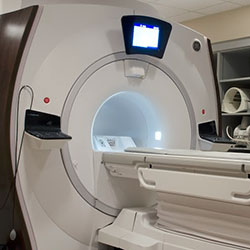
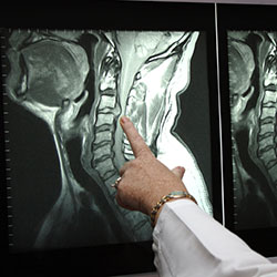
Magnetic resonance imaging (MRI) is a noninvasive, usually painless medical test that helps physicians diagnose and treat medical conditions.
MR imaging uses a powerful magnetic field, radio waves and a computer to produce detailed pictures of organs, soft tissues, bone and virtually all other internal body structures. The images can then be examined on a computer monitor or printed. MRI does not use ionizing radiation (x-rays).
Detailed MR images allow physicians to better evaluate parts of the body and certain diseases that may not be assessed adequately with other imaging methods such as x-ray,ultrasound or computed tomography (also called CT or CAT scanning).
For more information visit www.radiologyinfo.org.
Bring your insurance cards. Your insurance may require you to make a co-payment.
If you have had previous MRI studies from another facility not covered by the Radiological Group, please try to bring us a copy of that study and/or study report, if possible.
You should wear clothing without zippers and metal snaps. Jewelry such as watches, earrings, bracelets, and necklaces have to be taken off before entering MRI scanner area.
Follow the directions given for the location you are visiting found on our location pages. Upon arriving, check in with the receptionist who may have information for you to fill out. These forms are also available online. If you wish to fill them out ahead of time click here.
Once all your information has been obtained and processed, your technologist is notified of your arrival.
Your MRI technologist will talk with you before the exam, answering any questions you may have and asking you a series of screening questions to make sure you are a candidate for an MRI. People with electronic and metallic internal objects such as cardiac pace-makers, aneurysm clips, medication pumps, cochlear implants, etc. most often will not be able to have an MRI due to possible interactions between the magnetic and these devices. Click here for more information.
Some patients who have MRI in an enclosed scanner may feel confined, closed-in, and frightened. Perhaps one in twenty will require a sedative to remain calm. We have an “open” MRI at our Madison facility which is specifically designed to help those with claustrophobia. We will also permit a relative or friend to be present in the MR system room, which also has a calming effect. If patients are properly prepared and know what to expect, it is almost always possible to complete the examination.
You will placed on the MRI table. Once you are comfortable the table moves into the magnet. The MR machine makes a series of high volume sounds as it is acquiring images. You will be given ear plugs to help with the noise level. Alternatively, you can have earphones and listen to your favorite music during the study.
Depending on the type of MRI you are having, you may need intravenous or “IV” contrast which will be given through an IV line. The radiologist or technologist may ask if you have allergies of any kind such as hay fever, hives, allergic asthma, or to food or drugs. However, the contrast material used for an MRI exam, called gadolinium, does not contain iodine and is less likely to cause an allergic reaction.
One of the keys to an optimal study is to try to keep the body part being imaged as still as possible. For example, your knee for a MRI of the knee or your head for an MRI of the brain.
Study times vary based on the body part being imaged. Times may range from 20 to 45 minutes depending on the type of study. Times may be longer if your having multiple body parts imaged.
Any IV line will be removed. If you are undressed for the exam, you are taken back to the dressing room to put your clothes back on. Once the radiologist reviews your study and it is determined that no additional images or studies are needed, you will check out at the front desk. Depending on the order from your doctor, you will either stay while the report is given to your doctor or you will be free to leave and your doctor will discuss the study and results with you at a later time. All patients having an MRI are given a CD copy of their MRI study before leaving the facility.
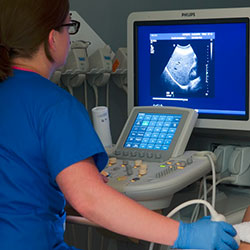
Ultrasound imaging, also called ultrasound scanning or sonography, involves exposing part of the body to high-frequency sound waves to produce pictures of the inside of the body. Ultrasound exams do not use ionizing radiation (x-ray). Because ultrasound images are captured in real-time, they can show the structure and movement of the body’s internal organs, as well as blood flowing through blood vessels.
Ultrasound imaging is usually a painless medical test that helps physicians diagnose and treat medical conditions.
Conventional ultrasound displays the images in thin, flat sections of the body. Advancements in ultrasound technology include three-dimensional (3-D) ultrasound that formats the sound wave data into 3-D images. Four-dimensional (4-D) ultrasound is 3-D ultrasound in motion.
A Doppler ultrasound study may be part of an ultrasound examination.
Doppler ultrasound is a special ultrasound technique that evaluates blood as it flows through a blood vessel, including the body’s major arteries and veins in the abdomen, arms, legs and neck.
For more information on ultrasound please visit radiologyinfo.org.
Abdomen or gallbladder ultrasound: Do not eat or drink after midnight (NPO), including no smoking or gum chewing. You may brush your teeth.
Pelvic Ultrasound: Do not go to the bathroom 2 hours before the exam. During the first hour before the exam, you need to drink one 8 ounce glass of clear liquid every 15 minutes. It can be any clear liquid (Kool-aid, coffee, coke, etc. Examples of nonclear liquids are milk or juice.) Stop drinking 1 hour before exam.
For example: If appointment time is 2:00 PM, do not go to the bathroom after 12:00 noon. Starting at 12 noon until 1:00 PM, drink one glass of water every 15 minutes. Stop drinking the water at 1:00 PM. Patient may come in early to complete paperwork, 15 minutes is usually enough time to complete the paperwork.
Abdomen and pelvic ultrasound: Do not eat after midnight. NO FOOD. Drink one 8 ounce glass of water every 15 minutes beginning 2 hours before appointment time. This will be a total of four 8 ounce glasses (32 ounces). Stop drinking one hour before your appointment time.
Renal ultrasound: You may eat or drink with no restrictions. Do not go to the bathroom one hour before the examination. You do not need to force fluids, just have a normally full bladder until the examination is completed.
All other types of ultrasounds: There are no preparations for other ultrasound examinations except for biopsies.
You need to bring your insurance card and driver’s license. You will not need a driver. You will not receive any medication.
You will be asked to remove most of your clothing and instructed to put on a gown. Sonograms require a gel be applied to your skin. This is so our transducer will glide across your skin and to insure there is contact between your skin and the transducer.
Follow the directions given for the location you are visiting found on our location pages. Upon arriving, check in with the receptionist who may have information for you to fill out. These forms are also available online. If you wish to fill them out ahead of time click here.
Once all your information has been obtained and processed, your technologist is notified of your arrival.
You will lie on an examination table. A warm gel will be applied to your skin, then a small instrument called a transducer will be moved across your skin. The transducer sends sound waves into your body. These high frequency sound waves are reflected back to the transducer. This information is processed by the ultrasound machine in real time producing sonographic images of the inside of your body. You cannot hear the sounds and the study is generally painless.
If you are female and are having a pelvic ultrasound, an endovaginal scan may be performed. This may be a little uncomfortable but should not be painful.
The most important thing is for you to follow the instructions for the preparation before the sonogram. During the study, you may be asked to take in a deep breath which helps move your internal organs away from bone and gas (as is in your stomach and colon). These structures can limit sonographic visualization.
Most examinations take 15-30 minutes.
If you are undressed for the exam, you are taken back to the dressing room to put your clothes back on. Once the radiologist reviews your study and it is determined that no additional images or studies are needed, you will check out at the front desk. Depending on the order from your doctor, you will either stay while the report is given to your doctor or you will be free to leave and your doctor will discuss the study and results with you at a later time.
The chest x-ray is the most commonly performed diagnostic x-ray examination. A chest x-ray produces images of the heart, lungs, airways, blood vessels and the bones of the spine and chest.
An x-ray (radiograph) is a noninvasive medical test that helps physicians diagnose and treat medical conditions. Imaging with x-rays involves exposing a part of the body to a small dose of ionizing radiation to produce pictures of the inside of the body. X-rays are the oldest and most frequently used form of medical imaging.
There are many other uses of XRay other than Chest. Please use this link to learn more:
http://www.radiologyinfo.org/en/sitemap/modal-alias.cfm?modal=xray
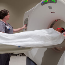
Computed Tomography (CT) System, also know as a “CAT Scan” is a medical diagnostic tool that allows the visualization of internal structures within the human body. This aids physicians in diagnosing disease, viewing internal abnormalities and assessing the extent of trauma damage.
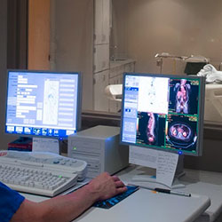
During a typical CT procedure, the patient is placed on a table. The table then moves the patient through the gantry (a donut-shaped device), which houses an X-ray tube and detector array. For each image acquired, the X-ray tube rotates around the patient and the X-rays pass through the patient to the detector array, and thousands of X-ray measurements are acquired. The computer then processes this information and displays the corresponding images on a computer screen. This imaging technique avoids any superimposition of organs or tissues upon one another that might occur during other types of X-ray tomographic studies.
The CT exam creates images analogous to a single slice of bread from a whole loaf or a slice from an orange. Hence, the word ‘slice’ is often used to describe a view of patient anatomy.
The quality of an image depends on the nature of the X-ray source and detectors, the number and speed of the measurements made, the details of the reconstruction technique (algorithm), the machine characteristics, and the methods of data display and interpretation. The computer allows healthcare professionals to shade, rotate, correlate and measure anatomy in the image. This data can be manipulated to derive even more precise clinical information. While conventional X-ray can discern tissue density difference of five percent, CT can distinguish a density difference of 1 percent or less, aiding in diagnostic confidence.
For more information, visit RSNA’s radiologyinfo.org website.
If you are having a CT of your abdomen and/or pelvis, you might have to drink oral contrast material which is given to coat the intestines. This is usually given before the exam. Therefore, we ask that you have nothing to eat or drink after midnight on the night before your exam.
Depending on the type of CT you are having, you may need intravenous or “IV” contrast which will be given through an IV line immediately before the study. IV contrast is given to enhance the blood vessels that feed your organs. We ask that you do not eat a heavy meal before your exam as, occasionally, patients receiving IV contrast who have had a heavy meal may experience mild nausea. During the study, IV contrast may make you feel warm all over. This can last for about 1 minute and is normal.
Please let us know ahead of time if you have a history of kidney disease or renal failure. If this is the case, you may need a blood test to determine your kidney function if you are to receive IV contrast.
If you are having a CT that requires IV contrast and are taking a medication for diabetes known as Metformin (Glucophage, Glucovance, etc.), you will need to stop taking this medication during the two days following your exam. Also, your doctor may need to order labwork to check your renal function. Your doctor will be aware what to do in this event.
We recommend drinking plenty of fluids after your exam to flush both the IV and oral contrast from your system.
Bring your insurance cards. Your insurance may require you to make a co-payment.
If you have had previous CT studies from another facility not covered by the Radiological Group, please try to bring us a copy of that study and/or study report, if possible.
We recommend wearing comfortable clothes that do not contain any metal in the area we are going to be imaging. We have gowns for you to change into in cases where this is needed.
Follow the directions given for the location you are visiting found on our location pages. Upon arriving, check in with the receptionist who may have information for you to fill out. These forms are also available online. If you wish to fill them out ahead of time click here.
Once all your information has been obtained and processed, your technologist is notified of your arrival.
The technologist will take you to the CT suite and ask you a series of history questions that aid in the reading of your exam. If needed, you may be given a cup of oral contrast to drink. If IV contrast is needed, the technologist will start an IV line. Once everything is ready, the technologist will start your study. You may be given breathing instructions during your study if needed for your exam.
During a CT scan, the images are obtained in a matter of seconds. During the scan, you are asked to hold still and sometimes hold your breath for a few seconds. Following these instructions can improve the image quality of your study.
CT studies usually range from 5-10 minutes though may be longer if it is determined during your study that additional images are needed.
Any IV line will be removed. If you are undressed for the exam, you are taken back to the dressing room to put your clothes back on. Once the radiologist reviews your study and it is determined that no additional images or studies are needed, you will check out at the front desk. Depending on the order from your doctor, you will either stay while the report is given to your doctor or you will be free to leave and your doctor will discuss the study and results with you at a later time.
Upper gastrointestinal tract radiography, also called an upper GI, is an x-ray examination of the esophagus, stomach and first part of the small intestine (also known as the duodenum). Images are produced using a special form of x-ray called fluoroscopy and an orally ingested contrast material such as barium
An x-ray (radiograph) is a noninvasive medical test that helps physicians diagnose and treat medical conditions. Imaging with x-rays involves exposing a part of the body to a small dose of ionizing radiation to produce pictures of the inside of the body. X-rays are the oldest and most frequently used form of medical imaging.
Fluoroscopy makes it possible to see internal organs in motion. When the upper GI tract is coated with barium, the radiologist is able to view and assess the anatomy and function of the esophagus, stomach and duodenum.
An x-ray examination that evaluates only the pharynx and esophagus is called a barium swallow.
In addition to drinking barium, some patients are also given baking-soda crystals (similar to Alka-Seltzer) to further improve the images. This procedure is called an air-contrast or double-contrast upper GI.
On occasion, some patients are given other forms of orally ingested contrast, usually containing iodine. These alternative contrast materials may be used if the patient has recently undergone surgery on the GI tract, or has allergies to other contrast materials. The radiologist will determine which type of contrast material will be used.
An upper GI examination helps evaluate digestive function and can detect:
The procedure is also used to help diagnose the cause of symptoms such as:
Your physician will give you detailed instructions on how to prepare for your upper GI.
You should inform your physician of any medications being taken and if there are any allergies, especially to iodinated contrast materials. Also inform your doctor about recent illnesses or other medical conditions.
Women should always inform their physician and x-ray technologist if there is any possibility that they are pregnant. Many imaging tests are not performed during pregnancy so as not to expose the fetus to radiation. If an x-ray is necessary, precautions will be taken to minimize radiation exposure to the baby. See the Safety page for more information about pregnancy and x-rays.
To ensure the best possible image quality, your stomach must be empty of food. Therefore, your doctor will likely ask you not to eat or drink anything (including any medications taken by mouth, especially antacids) and to refrain from chewing gum after midnight on the day of the examination.
You may be asked to remove some or all of your clothes and to wear a gown during the exam. You may also be asked to remove jewelry, removable dental appliances, eye glasses and any metal objects or clothing that might interfere with the x-ray images.
The equipment typically used for this examination consists of a radiographic table, one or two x-ray tubes and a television-like monitor that is located in the examining room. Fluoroscopy, which converts x-rays into video images, is used to watch and guide progress of the procedure. The video is produced by the x-ray machine and a detector that is suspended over a table on which the patient lies.
X-rays are a form of radiation like light or radio waves. X-rays pass through most objects, including the body. Once it is carefully aimed at the part of the body being examined, an x-ray machine produces a small burst of radiation that passes through the body, recording an image on photographic film or a special detector.
Fluoroscopy uses a continuous or pulsed x-ray beam to create a sequence of images that are projected onto a fluorescent screen, or television-like monitor. When used with a contrast material, which clearly defines the area being examined by making it appear dark (or by electronically reversing the image contrast to white), this special x-ray technique makes it possible for the physician to view joints or internal organs in motion. Still images or movies are also captured and stored electronically on a computer.
Until recently, x-ray images were maintained as hard film copy (much like a photographic negative). Today, most images are digital files that are stored electronically. These stored images are easily accessible and are frequently compared to current x-ray images for diagnosis and disease management.
As the patient drinks the liquid barium, which resembles a light-colored milkshake, the radiologist will watch the barium pass through the patient’s digestive tract on a fluoroscope, a device that projects radiographic images in a movie-like sequence onto a monitor. The exam table will be positioned at different angles and the patient’s abdomen may be compressed to help spread the barium. Once the upper GI tract is adequately coated with the barium, still x-ray images will be taken and stored for further review.
Children usually drink barium contrast material without any objection. If a child will not drink the contrast, the radiologist may need to pass a small tube into the stomach to complete the examination.
Very young children may be placed on a special rotating platform to help turn them into slanted positions. This allows the radiologist to see all the organs. Older children will be asked to hold very still and may be asked to hold their breath for a few seconds while the x-ray pictures are taken.
Older children may undergo a double-contrast upper GI series. The patient will swallow baking-soda crystals that create gas in the stomach while additional x-rays are taken.
When the examination is complete, you will be asked to wait until the radiologist determines that all the necessary images have been obtained.
This exam is usually completed within 20 minutes.
Occasionally, patients find the thick consistency of the barium unpleasant and difficult to swallow. The liquid barium has a chalky taste that may be masked somewhat by added flavors such as strawberry or chocolate.
Being tilted on the examination table and having pressure applied to the abdomen can be uncomfortable for some patients. The examination may also make you feel bloated.
If you receive gas-producing crystals, you may feel the need to belch. However, the radiologist or technologist will tell you to try to hold the gas in (by swallowing your saliva if necessary) to enhance the detail on the x-ray images.
In some medical centers, the technologist can minimize patient movement by automatically tilting the examining table. These actions ensure that the barium is coating all parts of the upper GI tract. As the procedure continues, the technologist or the radiologist may ask you to drink more barium. You may hear the mechanical noises of the radiographic apparatus moving into place during the exam.
After the examination, you can resume a regular diet and take orally administered medications unless instructed otherwise by your doctor.
The barium may color your stools gray or white for 48 to 72 hours after the procedure. Sometimes the barium can cause temporary constipation, which is usually treated by an over-the-counter laxative. Drinking large quantities of fluids for several days following the test can also help. If you are unable to have a bowel movement or if your bowel habits undergo any significant changes following the exam, you should contact your physician.
A radiologist, a physician specifically trained to supervise and interpret radiology examinations, will analyze the images and send a signed report to your primary care or referring physician, who will discuss the results with you.
Follow-up examinations may be necessary, and your doctor will explain the exact reason why another exam is requested. Sometimes a follow-up exam is done because a suspicious or questionable finding needs clarification with additional views or a special imaging technique. A follow-up examination may also be necessary so that any change in a known abnormality can be monitored over time. Follow-up examinations are sometimes the best way to see if treatment is working or if an abnormality is stable over time.
Special care is taken during x-ray examinations to use the lowest radiation dose possible while producing the best images for evaluation. National and international radiology protection organizations continually review and update the technique standards used by radiology professionals.
Modern x-ray systems have very controlled x-ray beams and dose control methods to minimize stray (scatter) radiation. This ensures that those parts of a patient’s body not being imaged receive minimal radiation exposure.
See more information at thttp://www.radiologyinfo.org/en/info.cfm?pg=uppergi
Lower gastrointestinal (GI) tract radiography, also called a lower GI or barium enema, is an x-ray examination of the large intestine, also known as the colon. This examination evaluates the right or ascending colon, the transverse colon, the left or descending colon, the sigmoid colon and the rectum. The appendix and a portion of the distal small intestine may also be included.
An x-ray (radiograph) is a noninvasive medical test that helps physicians diagnose and treat medical conditions. Imaging with x-rays involves exposing a part of the body to a small dose of ionizing radiationto produce pictures of the inside of the body. X-rays are the oldest and most frequently used form of medical imaging.
The lower GI uses a special form of x-ray called fluoroscopy and a contrast material called barium or a water soluble iodinated contrast.
Fluoroscopy makes it possible to see internal organs in motion. When the lower gastrointestinal tract is filled with barium, theradiologist is able to view and assess the anatomy and function of the rectum, colon and sometimes part of the lower small intestine.
A physician may order a lower GI examination to detect:
Images of the small bowel and colon are also used to diagnose inflammatory bowel disease, a group of disorders that includesCrohn’s disease and ulcerative colitis.
Your physician will give you detailed instructions on how to prepare for your lower GI imaging.
You should inform your physician of any medications being taken and if there are any allergies, especially to iodinated contrast materials. Also inform your doctor about recent illnesses or other medical conditions.
On the day before the procedure you will likely be asked not to eat, and to drink only clear liquids like juice, tea, black coffee, cola or broth, and to avoid dairy products. After midnight, you should not eat or drink anything. For adults (but not usually in children), it is important that your colon be completely empty for the procedure. You may also be instructed to take a laxative (in either pill or liquid form) and to use an over-the-counter enema preparation the night before the examination and possibly a few hours before the procedure. Just follow your doctor’s instructions. You can take your usual prescribed oral medications with limited amounts of water.
You may be asked to remove some or all of your clothes and to wear a gown during the exam. You may also be asked to remove jewelry, removable dental appliances, eye glasses and any metal objects or clothing that might interfere with the x-ray images.
Women should always inform their physician and x-ray technologist if there is any possibility that they are pregnant. Many imaging tests are not performed during pregnancy so as not to expose the fetus to radiation. If an x-ray is necessary, precautions will be taken to minimize radiation exposure to the baby. See the Safety page for more information about pregnancy and x-rays.
Infants and children may undergo lower GI radiography. Usually, there is no special preparation, but your doctor will give you detailed instructions to prepare your child for the examination. The use of barium and the taking of x-ray images is similar to that described for adults.
The equipment typically used for this examination consists of a radiographic table, one or two x-ray tubes and a television-like monitor that is located in the examining room. Fluoroscopy, which converts x-rays into video images, is used to watch and guide progress of the procedure. The video is produced by the x-ray machine and a detector that is suspended over a table on which the patient lies.
X-rays are a form of radiation like light or radio waves. X-rays pass through most objects, including the body. Once it is carefully aimed at the part of the body being examined, an x-ray machine produces a small burst of radiation that passes through the body, recording an image on photographic film or a special detector.
Fluoroscopy uses a continuous or pulsed x-ray beam to create a sequence of images that are projected onto a fluorescent screen, or television-like monitor. When used with a contrast material, which clearly defines the area being examined by making it appear dark (or by electronically reversing the image contrast to white), this special x-ray technique makes it possible for the physician to view joints or internal organs in motion. Still images or movies are also captured and stored electronically on a computer.
Until recently, x-ray images were maintained as hard film copy (much like a photographic negative). Today, most images are digital files that are stored electronically. These stored images are easily accessible and are frequently compared to current x-ray images for diagnosis and disease management.
The lower GI examination is usually done on an outpatient basis and is often scheduled in the morning to reduce the patient’s fasting time.
A radiology technologist and a radiologist, a physician specifically trained to supervise and interpret radiology examinations, guide the patient through the barium enema.
The patient is positioned on the examination table and an x-ray film is taken to ensure the bowel is clean. After performing a rectal examination, the radiologist or technologist will then insert a small tube into the rectum and begin to instill, using gravity, a mixture of barium and water into the colon. Air may also be injected through the tube to help the barium thoroughly coat the lining of the colon. In some circumstances, the radiologist or referring physician may prefer a water and iodine solution rather than barium. Next, a series of x-ray images is taken.
You must hold very still and may be asked to keep from breathing for a few seconds while the x-ray picture is taken to reduce the possibility of a blurred image. The technologist will walk behind a wall or into the next room to activate the x-ray machine.
The patient may be repositioned frequently in order to image the colon from several angles. Some x-ray equipment will allow patients to remain in the same position throughout the examination.
When the examination is complete, you will be asked to wait until the radiologist determines that all the necessary images have been obtained.
Once the x-ray images are completed, most of the barium will be emptied through the tube. The patient will then expel the remaining barium and air in the restroom. In some cases, additional x-ray images will be taken.
A barium enema is usually completed within 30 to 60 minutes.
As the barium fills your colon, you will feel the need to move your bowel. You may feel abdominal pressure or even minor cramping. Most people tolerate the mild discomfort easily. The tip of the enema tube is specially designed to help you hold in the barium. If you are having trouble, let the technologist or radiologist know.
During the imaging process, you will be asked to turn from side to side and to hold several different positions. At times, pressure may be applied to your abdomen. With air contrast studies of the bowel (air contrast barium enema), the table may be moved to an upright position.
After the examination, you may be given a laxative or enema to wash the barium out of your system. You can resume a regular diet and take orally administered medications unless told otherwise by your doctor. You may be able to return to a normal diet and activities immediately after the examination. You will be encouraged to drink additional water for 24 hours after the examination.
Your stools may appear white for a day or so as your body clears the barium liquid from your system. Some people experience constipation after a barium enema. If you do not have a bowel movement for more than two days after your exam or are unable to pass gas rectally, call your physician promptly. You may need an enema or laxative to assist in eliminating the barium.
A radiologist, a physician specifically trained to supervise and interpret radiology examinations, will analyze the images and send a signed report to your primary care or referring physician, who will discuss the results with you.
Follow-up examinations may be necessary, and your doctor will explain the exact reason why another exam is requested. Sometimes a follow-up exam is done because a suspicious or questionable finding needs clarification with additional views or a special imaging technique. A follow-up examination may also be necessary so that any change in a known abnormality can be monitored over time. Follow-up examinations are sometimes the best way to see if treatment is working or if an abnormality is stable over time.
Special care is taken during x-ray examinations to use the lowest radiation dose possible while producing the best images for evaluation. National and international radiology protection organizations continually review and update the technique standards used by radiology professionals.
Modern x-ray systems have very controlled x-ray beams and dose control methods to minimize stray (scatter) radiation. This ensures that those parts of a patient’s body not being imaged receive minimal radiation exposure.
Get more information at http://www.radiologyinfo.org/en/info.cfm?pg=lowergi
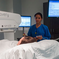
Nuclear medicine is a branch of medical imaging that uses small amounts of radioactive material to diagnose and determine the severity of or treat a variety of diseases, including many types of cancers, heart disease, gastrointestinal, endocrine, neurological disorders and other abnormalities within the body. Because nuclear medicine procedures are able to pinpoint molecular activity within the body, they offer the potential to identify disease in its earliest stages as well as a patient’s immediate response to therapeutic interventions.
For more information: http://www.radiologyinfo.org/en/info.cfm?PG=gennuclear
Positron emission tomography, also called PET imaging or a PET scan, is a type of nuclear medicine imaging.
Nuclear medicine is a branch of medical imaging that uses small amounts of radioactive material to diagnose and determine the severity of or treat a variety of diseases, including many types of cancers, heart disease, gastrointestinal, endocrine, neurological disorders and other abnormalities within the body. Because nuclear medicine procedures are able to pinpoint molecular activity within the body, they offer the potential to identify disease in its earliest stages as well as a patient’s immediate response to therapeutic interventions.
Nuclear medicine imaging procedures are noninvasive and, with the exception of intravenous injections, are usually painless medical tests that help physicians diagnose and evaluate medical conditions. These imaging scans use radioactive materials calledradiopharmaceuticals or radiotracers.
Depending on the type of nuclear medicine exam, the radiotracer is either injected into the body, swallowed or inhaled as a gas and eventually accumulates in the organ or area of the body being examined. Radioactive emissions from the radiotracer are detected by a special camera or imaging device that produces pictures and provides molecular information.
In many centers, nuclear medicine images can be superimposed with computed tomography (CT) ormagnetic resonance imaging (MRI) to produce special views, a practice known as image fusion or co-registration. These views allow the information from two different exams to be correlated and interpreted on one image, leading to more precise information and accurate diagnoses. In addition, manufacturers are now making single photon emission computed tomography/computed tomography (SPECT/CT) and positron emission tomography/computed tomography (PET/CT) units that are able to perform both imaging exams at the same time. An emerging imaging technology, but not readily available at this time is PET/MRI.
A PET scan measures important body functions, such as blood flow, oxygen use, and sugar (glucose)metabolism, to help doctors evaluate how well organs and tissues are functioning.
CT imaging uses special x-ray equipment, and in some cases a contrast material, to produce multiple images or pictures of the inside of the body. These images can then be interpreted by a radiologist on a computer monitor. CT imaging provides excellent anatomic information.
Today, almost all PET scans are performed on instruments that are combined PET and CT scanners. The combined PET/CT scans provide images that pinpoint the anatomic location of abnormal metabolic activity within the body. The combined scans have been shown to provide more accurate diagnoses than the two scans performed separately.
PET and PET/CT scans are performed to:
You may be asked to wear a gown during the exam or you may be allowed to wear your own clothing.
Women should always inform their physician or technologist if there is any possibility that they are pregnant or if they are breastfeeding. See the Safety page for more information about pregnancy and breastfeeding related to nuclear medicine imaging.
You should inform your physician and the technologist performing your exam of any medications you are taking, including vitamins and herbal supplements. You should also inform them if you have any allergies and about recent illnesses or other medical conditions.
You will receive specific instructions based on the type of PET scan you are undergoing. Diabetic patients will receive special instructions to prepare for this exam.
If you are breastfeeding at the time of the exam, you should ask your radiologist or the doctor ordering the exam how to proceed. It may help to pump breast milk ahead of time and keep it on hand for use after the PET radiopharmaceutical and CT contrast material are no longer in your body.
Metal objects including jewelry, eyeglasses, dentures and hairpins may affect the CT images and should be left at home or removed prior to your exam. You may also be asked to remove hearing aids and removable dental work.
Generally, you will be asked not to eat anything for several hours before a whole body PET/CT scan since eating may alter the distribution of the PET tracer in your body and can lead to a suboptimal scan. This could require the scan to be repeated on another day, so following instructions regarding eating is very important. You should not drink any liquids containing sugars or calories for several hours before the scan. Instead, you are encouraged to drink water. If you are diabetic, you may be given special instructions. You should inform your physician of any medications you are taking and if you have any allergies, especially to contrast materials, iodine, or seafood.
You will be asked and checked for any conditions that you may have that may increase the risk of receiving intravenous contrast material.
A PET scanner is a large machine with a round, doughnut shaped hole in the middle, similar to a CT or MRI unit. Within this machine are multiple rings of detectors that record the emission of energy from the radiotracer in your body.
The CT scanner is typically a large, box-like machine with a hole, or short tunnel, in the center. You will lie on a narrow examination table that slides into and out of this tunnel. Rotating around you, the x-ray tube and electronic x-ray detectors are located opposite each other in a ring, called a gantry. The computer workstation that processes the imaging information is located in a separate control room, where the technologist operates the scanner and monitors your examination in direct visual contact and usually with the ability to hear and talk to you with the use of a speaker and microphone.
Combined PET/CT scanners are combinations of both scanners and look similar to both the PET and CT scanners.
A computer aids in creating the images from the data obtained by the gamma camera.
With ordinary x-ray examinations, an image is made by passing x-rays through the patient’s body. In contrast, nuclear medicine procedures use a radioactive material, called a radiopharmaceutical or radiotracer, which is injected into the bloodstream, swallowed or inhaled as a gas. This radioactive material accumulates in the organ or area of your body being examined, where it gives off a small amount of energy in the form of gamma rays. Special cameras detect this energy, and with the help of a computer, create pictures offering details on both the structure and function of organs and tissues in your body.
Unlike other imaging techniques, nuclear medicine imaging exams focus on depicting physiologic processes within the body, such as rates of metabolism or levels of various other chemical activity, instead of showing anatomy and structure. Areas of greater intensity, called “hot spots,” indicate where large amounts of the radiotracer have accumulated and where there is a high level of chemical or metabolic activity. Less intense areas, or “cold spots,” indicate a smaller concentration of radiotracer and less chemical activity.
For more information on how a CT scan works, see Computed Tomography at (www.RadiologyInfo.org/en/sitemap/modal-alias.cfm?modal=CT).
Nuclear medicine imaging is usually performed on an outpatient basis, but is often performed on hospitalized patients as well.
You will be positioned on an examination table. If necessary, a nurse or technologist will insert an intravenous (IV) catheter into a vein in your hand or arm.
Depending on the type of nuclear medicine exam you are undergoing, the dose of radiotracer is then injected intravenously, swallowed or inhaled as a gas.
Typically, it will take approximately 60 minutes for the radiotracer to travel through your body and to be absorbed by the organ or tissue being studied. You will be asked to rest quietly, avoiding movement and talking.
You may be asked to drink some contrast material that will localize in the intestines and help the radiologist interpreting the study.
You will then be moved into the PET/CT scanner and the imaging will begin. You will need to remain still during imaging. The CT exam will be done first, followed by the PET scan. On occasion, a second CT scan with intravenous contrast will follow the PET scan. For more information on how a CT scan is performed, see Computed Tomography at(www.RadiologyInfo.org/en/sitemap/modal-alias.cfm?modal=CT). The actual CT scanning takes less than two minutes. The PET scan takes 20-30 minutes.
Total scanning time is approximately 30 minutes.
Depending on which organ or tissue is being examined, additional tests involving other tracers or drugs may be used, which could lengthen the procedure time to three hours. For example, if you are being examined for heart disease, you may undergo a PET scan both before and after exercising or before and after receiving intravenous medication that increases blood flow to the heart.
When the examination is completed, you may be asked to wait until the technologist checks the images in case additional images are needed. Occasionally, more images are obtained for clarification or better visualization of certain areas or structures. The need for additional images does not necessarily mean there was a problem with the exam or that something abnormal was found, and should not be a cause of concern for you.
If you had an intravenous line inserted for the procedure, it will usually be removed unless you are scheduled for an additional procedure that same day that requires an intravenous line.
Except for intravenous injections, most nuclear medicine procedures are painless and are rarely associated with significant discomfort or side effects.
When the radiotracer is given intravenously, you will feel a slight pin prick when the needle is inserted into your vein for the intravenous line. When the radioactive material is injected into your arm, you may feel a cold sensation moving up your arm, but there are generally no other side effects.
When swallowed, the radiotracer has little or no taste. When inhaled, you should feel no differently than when breathing room air or holding your breath.
With some procedures, a catheter may be placed into your bladder, which may cause temporary discomfort.
It is important that you remain still while the images are being recorded. Though nuclear imaging itself causes no pain, there may be some discomfort from having to remain still or to stay in one particular position during imaging.
If you are claustrophobic, you may feel some anxiety while you are being scanned.
Unless your physician tells you otherwise, you may resume your normal activities after your nuclear medicine scan. If any special instructions are necessary, you will be informed by a technologist, nurse or physician before you leave the nuclear medicine department.
Through the natural process of radioactive decay, the small amount of radiotracer in your body will lose its radioactivity over time. It may also pass out of your body through your urine or stool during the first few hours or days following the test. You should also drink plenty of water to help flush the radioactive material out of your body as instructed by the nuclear medicine personnel.
For more information on what you will experience during and after a CT scan, see Computed Tomography at(www.RadiologyInfo.org/en/sitemap/modal-alias.cfm?modal=CT).